Warning: This article includes descriptions and images of a surgical procedure for mass removal.
A 10-year-old neutered male Australian Shepherd mix was presented for a second opinion after being diagnosed with a possible Hemangiosarcoma. Hemangiosarcoma is a fast-growing, highly invasive variety of cancer that arises from the blood vessels. If this were the case options would include surgery (which could cost up to $5,000.00) or euthanasia. At first, it seemed as if the growth was attached to the spleen and bladder, but the ultrasound implied it was only attached to the spleen. Doctor Neumeister did an exploratory laparotomy (a surgical operation where the abdomen is opened and the abdominal organs examined for injury or disease). The growth was even bigger than expected and it was confirmed to be only attached to the spleen. After speaking to the dog’s owners it was decided to remove the spleen with the growth and send a specimen to a specialty lab for a histopath.
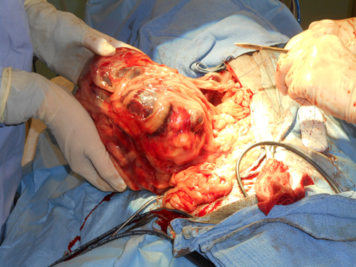 A Veterinary Technician is holding the growth while Dr. Neumeister is tying off blood vessels before removing the growth. |
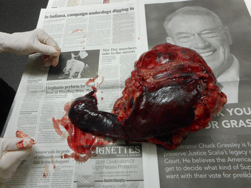
The spleen with the attached mass.
The mass weighed 5 pounds!
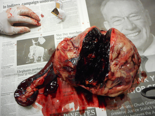
The lab results came back after 4 days and showed that the growth was a splenic hematoma (injury to a blood vessel wall of the spleen, prompting blood to seep out of the blood vessel into the surrounding tissues) and no evidence of malignancy was seen. The dog had regained it’s energy and strength within days and is still in good health today.