A client came to the office with her 10-year-old Boston Terrier because she felt that his vision was impaired.
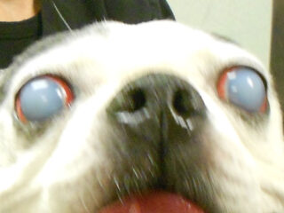 Dr. Neumeister measured the fluid pressure inside the eye (intraocular pressure) and evaluated the retina and optic nerve function with color-based pupil testing. Both tests came back with results in the normal range. After ruling out other eye diseases corneal endothelial dystrophy was diagnosed.
Corneal endothelial dystrophy is an age-related degeneration of the inner cell layer (endothelium) of the cornea (the cornea is a clear “window” that covers the front of the eye) which results in fluid movement into the corneal layers causing the opacification of the cornea. The edema/opacification usually starts temporal (outside margin of the eye) and progresses axial (toward the central aspect and nose). The result may be complete bilateral edema/opacification. Bullae, ulcers, and corneal vascularization may develop. Vision can be impaired in advanced cases. The condition is progressive and treatment is directed toward palliation/relief of the symptoms. Treatment is not curative.
This disease can be found in older dogs in any breed but the highest prevalence is in the Basset Hound, Boston Terrier, Chihuahua, Cocker Spaniel, Dachshund, English Springer Spaniel, West Highland White Terrier, and Wire Fox Terrier. The age of onset is five to nine years.